Use of Implantable Gold Fiducial Markers in Radiation Therapy
The use of implantable gold fiducial markers in the planning of radiation has become a viable option to accurately deliver external beam radiation to a patient’s treatment site.
Treating certain areas such as the prostate, lungs, breast, liver, etc., have been shown to move during treatment with respect to internal bony or external markings. Gold fiducial markers are implantable markers used during IMRT (Intensity Modulated Radiation Therapy) and IGRT (Image Guided Radiation Therapy) treatments to provide real-time localization of these moving volumes. Because soft tissues, like those listed above, are not visible or clearly distinguishable from the surrounding tissues within the body under normal imaging, this gives a valued reason to implant gold markers within these tissues.
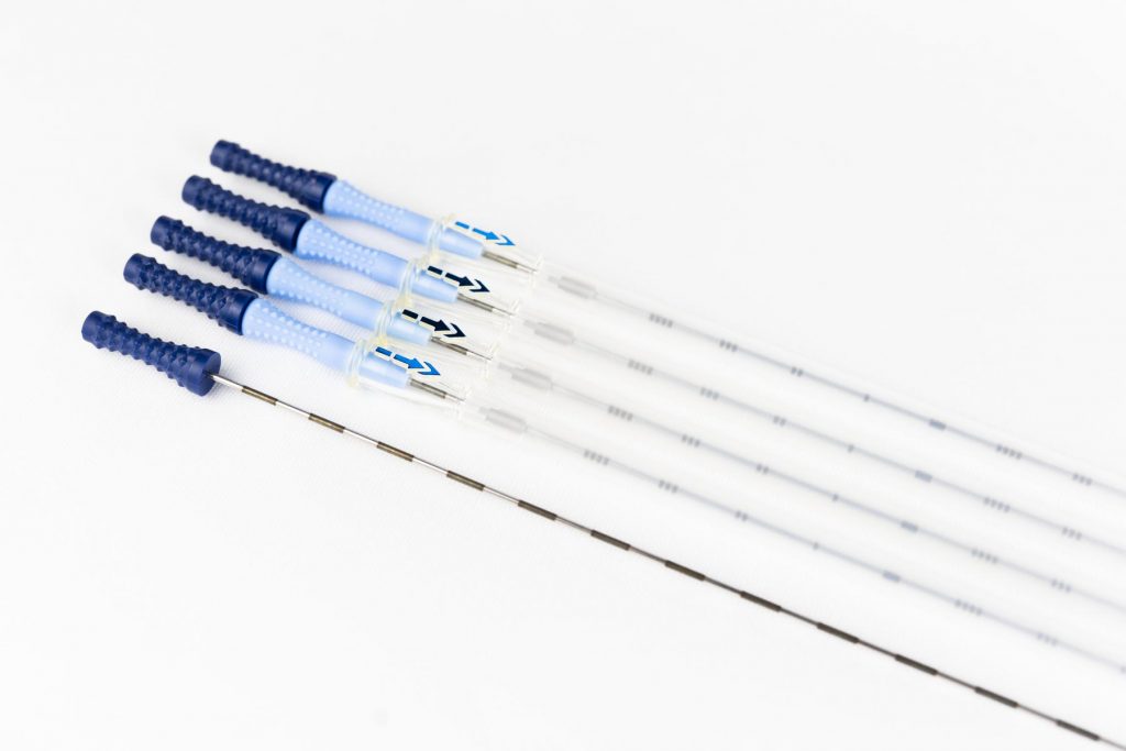
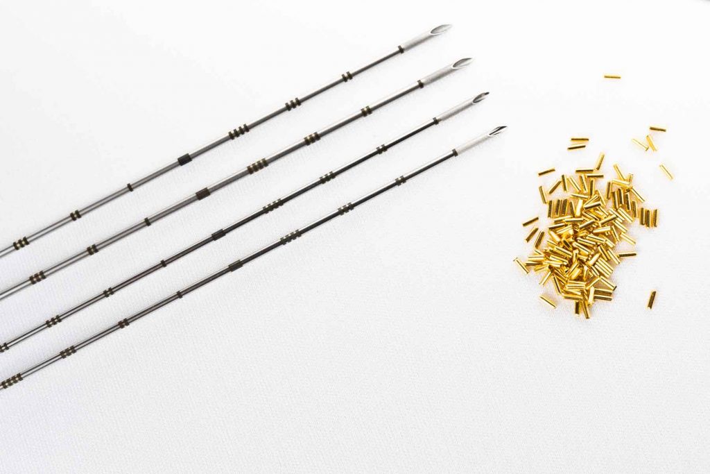
Gold markers are implanted by an experienced physician within that specialty like that of a Urologist. This procedure is considered a non-invasive outpatient procedure. The gold markers are implanted via sterilized needles under the guidance of a variety of imaging methods such as ultrasound or CT. A few days after implantation, the patient is scheduled for a CT scan for radiation treatment planning. The CT scan obtained will show the gold markers that were implanted within the area of concern. The number of markers implanted will vary by institution.
The CT images are reviewed by the radiation oncologist. With the assistance of a dosimetrist or medical physicist, the gold markers are delineated by digitally contouring them within the treatment planning computer system. These markers are easily seen on a CT image due to the fact that metal objects show up as a solid white artifact. Treatment planning is completed with a sufficient amount of radiation beams to provide an adequate isodose distribution to cover the tumor volume. An IMRT treatment plan is generated and approved by the prescribing Radiation Oncologist.
When the patient returns to the Radiation Therapy Department for his or her external beam radiation, they are placed on the treatment table under the linear accelerator. External markings are used to initially align the patient to the area of concern. The Radiation Therapist will then acquire an image of the patient in treatment position through the use of an OBI (On Board Imager). This image is generated using KV, MV or Cone-beam CT imaging techniques. The reconstructed image is shown on an image reconstructor computer and superimposed on the original CT image that contains the delineated gold markers. At this point, the Therapist can adjust the patient’s physical position based on the real-time location of the gold markers within the targeted area. The radiation beam can now be initialized and delivered with known accuracy. This is known as IGRT.
The use of implantable gold fiducial markers allow the treatment area of concern to be located before a radiation treatment is delivered. This improves the delivery and decreases the radiation exposure to the surrounding normal tissues.